Description
The surgeon makes small incisions (called portals) through the skin in specific locations around a joint.
These incisions are less than half an inch long. The arthroscope, which is approximately the size of a pencil, is inserted through these incisions. The arthroscope contains a small lens, a miniature camera, and a lighting system.
The three-dimensional images of the joint are projected through the camera onto a television monitor. The surgeon watches the monitor as he or she moves the instrument within the joint.
Probes, forceps, knives, and shavers at the ends of the arthroscope are used to correct problems uncovered by the surgeon.
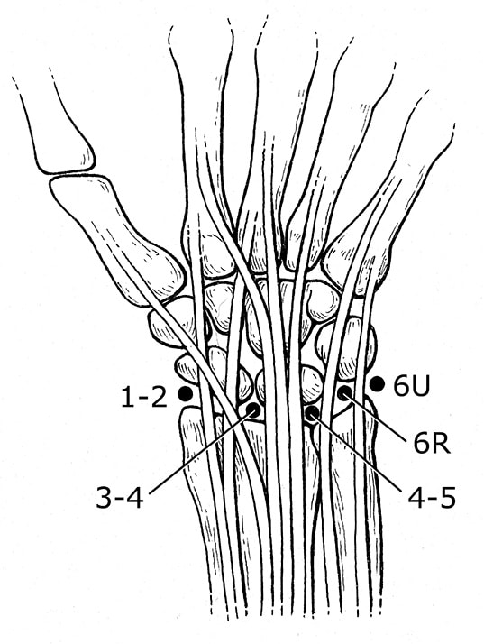
Surgeons place portals in specific locations on the wrist, depending on the area that needs to be viewed. This set of portals, called the standard radiocarpal portals, will give access to the wrist. Other sets of portals provide access to other areas of the wrist.
AnatomyhELLO
Arthroscopy is a surgical procedure used to diagnose and treat problems inside a joint.
Arthroscopy utilizes a small fiber optic instrument called an arthroscope that enables the surgeon to see inside the joint without making large incisions into the muscle and tissue.
The wrist is a complex joint with eight small bones and many connecting ligaments. Arthroscopic surgery can be used to diagnose and treat a number of conditions of the wrist, including chronic wrist pain, wrist fractures, ganglion cysts, and ligament tears.
Diagnostic Arthroscopy
Diagnostic arthroscopy might be used if it is not clear what is causing wrist pain. It might also be used if wrist pain continues for several months despite nonsurgical treatment.
Before arthroscopic surgery, your doctor will do the following.
- Perform a physical examination of the hand and wrist
- Learn more about past medical conditions or concerns (medical history)
- Perform tests that locate the pain (provocative tests). These tests involve moving the hand in order to reproduce the pain.
- Secure images of the hand and wrist. These may include x-rays, magnetic resonance imaging (MRI) scans, or an arthrogram (an x-ray taken after a dye is injected into the joint).
Usually, arthroscopic surgery requires only that the hand and arm are numbed (regional anesthesia). A sedative may be given to further relax the patient.
Two or more small incisions (portals) are made on the back of the wrist. The arthroscope and instruments are inserted through those portals and the joint is observed through the camera on the end of the arthroscope.
After the surgery, the incisions are closed with a small stitch and a dressing is applied. Sometimes a splint is used.
Treatment
Arthroscopic surgery can be used to treat a number of conditions of the wrist.
Surgical Treatment
Arthroscopic surgery can be used to treat a number of conditions of the wrist.
- Chronic wrist pain. Arthroscopic exploratory surgery may be used to diagnose the cause of chronic wrist pain when the results of other tests do not provide a clear diagnosis. Often, there may be areas of inflammation, cartilage damage, or other findings after a wrist injury. In some cases, after the diagnosis is made, the condition can be treated arthroscopically as well.
- Wrist fractures. Small fragments of bone may stay within the joint after a bone breaks (fractures). Wrist arthroscopy can remove these fragments, align the broken pieces of bone, and stabilize them by using pins, wires, or screws.
- Ganglion cysts. Ganglion cysts commonly grow from a stalk between two of the wrist bones. During an arthroscopic procedure, the surgeon can remove the stalk, which may reduce the change that these cysts will return.
- Ligament/TFCC tears. Ligaments are fibrous bands of connective tissue that link or hinge bones. They provide stability and support to the joints. The TFCC is a cushioning structure within the wrist. A fall on an outstretched hand can tear ligaments, the TFCC, or both. The result is pain with movement or a clicking sensation. During arthroscopic surgery, the surgeon can repair the tears.
- Carpal tunnel release. Carpal tunnel syndrome is characterized by numbness or tingling in the hand, and sometimes with pain up the arm. It is caused by pressure on a nerve that passes through the carpal tunnel. (The carpal tunnel is formed by the wrist bones and a thick tissue roof.) Pressure can build up within the tunnel for many reasons, including irritation and swelling of the tissue (synovium) that covers the tendons. If the carpal tunnel syndrome does not respond to nonsurgical treatment, one option is to repair the area surgically. The surgeon would cut the ligament roof and enlarge the tunnel. This would reduce pressure on the nerve and relieve symptoms. This can sometimes be done using an arthroscope.
After Surgery
Small fragments of bone may stay within the joint after a bone breaks (fractures). Wrist arthroscopy can remove these fragments, align the broken pieces of bone, and stabilize them by using pins, wires, or screws.
For the first 2 or 3 days after surgery, the wrist should be elevated and the bandage should be kept clean and dry. Ice may help keep swelling down. There are exercises that can be used to help maintain motion and rebuild your strength. Although pain after surgery is usually mild, analgesic medications will help relieve any pain.

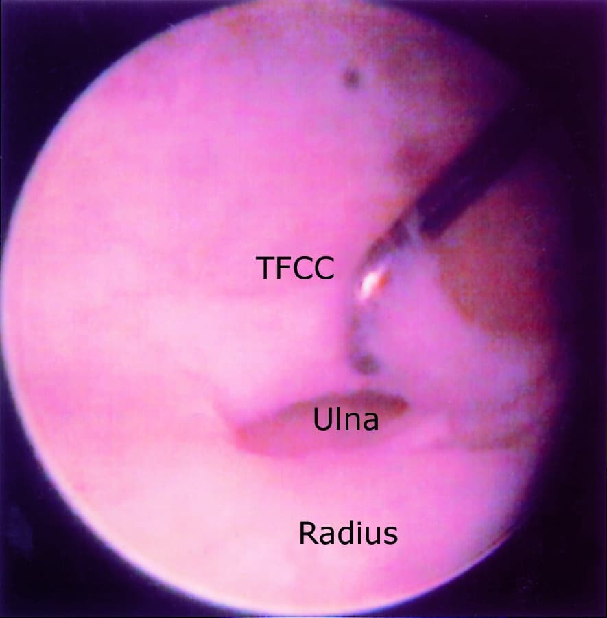
This is a view inside a joint using an arthroscope. The TFCC ligament is torn and the surgeon is using a hook to clean the area to make a smooth surface to suture the torn ligament in place. The head of the ulna is visualized through the tear. Next to it is the radius.
Reproduced from Gupta R, Bozentka DJ, Osterman AL: Wrist Arthroscopy: Principles and Clinical Applications. J. Am Acad. Orthop 2001;9:200-209.
ComplicationsS
Complications during or after arthroscopic wrist surgery are unusual. They may include infection, nerve injuries, excessive swelling, bleeding, scarring, or tendon tearing. Your doctor will discuss the complications of arthroscopy with you before your surgery.

