
Description
A scaphoid fracture is usually described by its location within the bone. Most commonly, the scaphoid breaks in its mid-portion, called the “waist.” Fractures can also occur at both the proximal and distal ends of the bone.
Scaphoid fractures are classified according to the severity of displacement–or how far the pieces of bone have moved out of their normal position:
- Non-displaced fracture. In this type of fracture, the bone fragments line up correctly.
- Displaced fracture. In this type of fracture, the bone fragments have moved out of their normal position. There may be gaps between the pieces of bone or fragments may overlap each other.


AnatomyhE
The wrist is formed by the two bones of the forearm—the radius and the ulna—and eight small carpal bones. The carpal bones are arranged in two rows at the base of the hand. There are four bones in each row.
The scaphoid bone is one of the carpal bones on the thumb side of the wrist, just above the radius. The bone is important for both motion and stability in the wrist joint. The word “scaphoid” comes from the Greek term for “boat.” The scaphoid bone resembles a boat with its relatively long, curved shape.
The scaphoid bone can most easily be identified when your thumb is held in a “hitch-hiking” position. It is located at the base of the hollow made by the thumb tendons. Often referred to as the “anatomic snuffbox,” this area is typically the site of tenderness or pain when a fracture occurs.


Figure (A): Normal anatomy of the hand and wrist. The scaphoid is one of the small carpal bones in the wrist.
Figure (B): Photograph and x-ray showing the location of the scaphoid in the wrist. The red arrows indicate the location of the anatomic snuffbox.
CausehE
A scaphoid fracture usually occurs when you fall onto an outstretched hand, with your weight landing on your palm. The end of the larger forearm bone (the radius) may also break in this type of fall, depending on the position of the hand on landing.
The injury can also happen during sports activities or motor vehicle collisions.
Fractures of the scaphoid occur in people of all ages, including children. There are no specific risk factors or diseases that make you more likely to experience a scaphoid fracture. Some studies have shown that using wrist guards during high-energy activities like inline skating and snowboarding can help decrease your chance of breaking a bone around the wrist.
SymptomshE
Scaphoid fractures usually cause pain and swelling in the anatomic snuffbox and on the thumb side of the wrist. The pain may be severe when you move your thumb or wrist, or when you try to pinch or grasp something.
Unless your wrist is deformed, it might not be obvious that your scaphoid bone is broken. With some scaphoid fractures, the pain is not severe and may be mistaken for a wrist sprain.
Pain in your wrist that does not go away within a day of injury may be a sign of a fracture—so it is important to see a doctor if your pain persists. Prompt treatment of a scaphoid fracture will help avoid potential complications.

Figure: Symptoms of a scaphoid fracture often occur in the anatomic snuffbox at the base of the thumb.
Doctor Examination
Physical Examination
During the exam, your doctor will talk with you about your general health and will ask you to describe your symptoms. He or she will want to know how your injury occurred.
Your doctor will examine your wrist. With most fractures, there will be tenderness directly over the scaphoid in the anatomic snuffbox. Your doctor will also look for:
- Swelling
- Bruising
- Loss of motion
Tests
- X-rays:
X-rays provide images of dense structures, such as bone. Your doctor will order an x-ray to help determine if you have a scaphoid fracture and whether the broken pieces of bone are displaced. An x-ray will also help your doctor determine if you have any other fractures.
In some cases, a scaphoid fracture does not show up on an x-ray right away. If your doctor suspects that you have a fracture but it is not visible on x-ray, he or she may recommend that you wear a wrist splint or cast for 2 to 3 weeks and then return for a follow-up x-ray. Often, scaphoid fractures become visible on x-ray only after a period of time. During this waiting period, you should wear your splint or cast and avoid activities that might cause further injury. - Magnetic resonance imaging (MRI) scan:
Your doctor may order an MRI to learn more about the bones and soft tissues in your wrist. An MRI can sometimes show a fracture of the scaphoid before it can be seen on x-ray. - Computerized tomography (CT) scan:
A CT scan can be helpful in revealing a fracture of the scaphoid and can also show whether the bones are displaced. Your doctor will use information from the CT scan to help determine your treatment plan.
Treatment
The treatment your doctor recommends will depend on a number of factors, including:
1. The location of the break in the bone 2. Whether the bone fragments are displaced. 3. How long ago your injury occurred
Nonsurgical Options
-
Fracture near the thumb:
Scaphoid fractures that are closer to the thumb (distal pole) usually heal in a matter of weeks with proper protection and restricted activity. This part of the scaphoid bone has a good blood supply, which is necessary for healing.
For this type of fracture, your doctor may place your forearm and hand in a cast or a splint. The cast or splint will usually be below the elbow and include your thumb.
Healing time varies from patient to patient. Your doctor will monitor your healing with periodic x-rays or other imaging studies. -
Fracture near the forearm:
If the scaphoid is broken in the middle of the bone (waist) or closer to the forearm (proximal pole), healing can be more difficult. These areas of the scaphoid do not have a very good blood supply.
If your doctor treats this type of fracture with a cast, the cast may include the thumb and extend above the elbow to help stabilize the fracture -
Bone stimulator:
In some cases, your doctor may recommend the use of a bone stimulator to assist in fracture healing. This small device delivers low-intensity ultrasonic or pulsed electromagnetic waves that stimulate healing.
Surgical Options
If your scaphoid is broken at the waist or proximal pole or if pieces of bone are displaced, your doctor may recommend surgery. The goal of surgery is to realign and stabilize the fracture, giving it a better chance to heal.
-
Reduction:
During this procedure, your doctor will administer an anesthetic or anesthesia and manipulate the bone back into its proper position. In some cases, this is done using a limited incision and special guided instruments. In other cases, it is performed through an open incision with direct manipulation of the fracture. For some fractures, your doctor may use a tiny camera called an “arthroscope” to aid in the reduction.
-
Internal fixation:
During this procedure, metal implants—including screws and/or wires—are used to hold the scaphoid in place until the bone is fully healed.
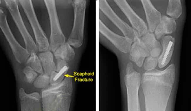
Figure: (Left) This x-ray shows a scaphoid fracture fixed in place with a screw. (Right) This x-ray was taken 4 months after surgery. The fracture of the scaphoid is healed.The location and size of the surgical incision depends on what part of the scaphoid is broken. Sometimes, the screw or wire can be placed in bone fragments with a small incision. In other cases, a larger incision is needed to ensure that the fragments of the scaphoid line up properly. The incision may be made on either the front or the back of your wrist.
-
Bone graft:
In some cases, a bone graft may be used with or without internal fixation. A bone graft is new bone that is placed around the broken bone. It can stimulate bone production and healing. The graft may be taken from your forearm bone in the same arm or from your hip.